
Patient Advisor
Welcome to the Patient Information Center. The information presented is intended to educate the general public on common dental topics. Many dental terms are commonly seen and heard in the print and broadcast media; as well as in our daily conversations. However, many people do not completely understand these dental terms unless they have had first hand experience with them at their dentist’s office.
This information is for educational purposes only and should not be used for self-diagnosis or as a substitute for a professional dental exam and consultation (no matter how much you hate going to the dentist). Each case will vary depending on the needs and concerns of each individual. We hope that this information will provide you with some basic dental knowledge so that you and your dentist can devise a plan to keep your smile healthy and looking great.
Having a clean mouth is important. In addition to being healthier, it gives you fresh breath and a nicer smile.
Are you Satisfied With Your Smile? Take A Small Test Now!!
Click on the below Headers to know more...
Brushing your teeth after meals and between-meal snacks not only gets rid of the food particles that you can see, it removes plaque from your teeth. Using a fluoride toothpaste is important because the fluoride can help kill bacteria, as well as make your teeth stronger.
Ask your dentist to recommend the best toothbrush for you. Generally, a brush with soft, end-rounded or polished bristles is less likely to injure gum tissue. The size and shape of the brush should allow you to reach every tooth. children may need smaller brushes than those designed for adults. Remember: worn-out toothbrushes can not properly clean your teeth and may injure your gums. Toothbrushes should be replaced every three or four months.
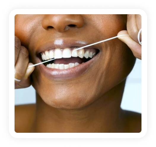
Flossing removes plaque and food particles from between teeth and under the gumline, areas your toothbrush can not reach. Because tooth decay and periodontal disease often start in these areas, it is important to clean them thoroughly on a daily basis.
Flossing is a skill that needs to be learned. Do not be discouraged if you find it difficult at first. With practice, you will find that flossing takes only a few minutes of your time each day.
What about mouthrinses and mouthwashes?
If used as directed, in addition to brushing and flossing, mouthrinses and mouthwashes can help to prevent tooth decay.
How often should I see my dentist?
If possible, you should visit your dentist every six months for a preventive check and cleaning. Infants should see a dentist at about 12 months of age.
Plaque
Research has shown that controlling plaque is important in the control of decay and gum disease. Plaque is neither food or food residue. Plaque is a clear, sticky deposit of of bacteria that adheres to the surface of teeth and gum tissue. It is so adherent that it can only be removed by mechanical cleansing. Plaque contains a variety of different types of bacteria. For this reason, certain types of plaque are associated with dental decay, others with calculus formation, and others with the inflammatory response of the gums (gingivitis).
Plaque begins forming on the teeth in as little as 4 hours after brushing. This is why it is so important to brush your teeth at least twice a day and floss daily. The rate at which plaque forms and the location in which it develops can vary between individuals and even between different teeth in the same mouth. One of the prime areas in which plaque accumulates is at the gingival margin and sulcus where the tooth meets the gum.
Calculus
Plaque which is not removed regularly by brushing and flossing can harden into calculus (also called tartar). Calculus is plaque that has mineralized, forming a tough, crusty deposit that can only be removed by your dentist or hygienist. These deposits can form above (supragingival) and below (subgingival) the gum line. Calculus deposits are a significant contributing factor in periodontal disease because it is always covered by a layer of nonmineralized plaque. The calculus keeps the plaque close to the gingival tissue and makes it much more difficult to remove the plaque bacteria. Thorough removal of these deposits is necessary to prevent the progression of periodontal disease.
Some people form heavy calculus deposits rapidly while others form little or no mineralized deposits. This is due to differences in the saliva, the types of plaque bacteria, and dietary factors. One can help reduce the formation of calculus by brushing with and ADA-accepted tartar control toothpaste and by having regular professional cleanings every 6 months or more frequently as recommended by your dentist or hygienist.
The prevention of gum disease and decay requires a life-long commitment to fighting plaque and calculus formation.
Sealants
Sealants are filling like materials that are placed on the chewing surfaces of permanent teeth to protect the grooves, pits and fissures from forming cavities.
Hard bristles were once recommended but are now thought to be too abrasive to the teeth and gums. We now suggest a soft, rounded-end nylon bristle brush. Be sure to discard brushes when the bristles are bent or frayed or approximately every three to four months.
How To Brush
Begin by placing the head of the brush beside your teeth, with the bristles angled against the gum line (where the teeth and gums meet ). Think of the brush as both a toothbrush and a gum brush. With the bristles contacting both tooth and gum, move the brush back and forth several times across each tooth individually.
Use a short stroke and a gentle scrubbing motion, as if the goal were to massage the gum. Don’t try to force the bristles under the gum line; that will happen naturally, especially with a brush that has soft, flexible bristles.
Brush the outer surfaces of the upper and lower teeth. Then use the same short back-and-forth strokes on the inside surfaces. Try to concentrate harder on the inside surfaces; studies show they’re more often neglected. For the upper and lower front teeth, brush the inside surfaces by using the brush vertically and making several gentle up–and-down strokes over the teeth and gums.
Finish up by lightly scrubbing the chewing surfaces of the upper and lower teeth. You should also brush your tongue for a fresher breath.
Flossing Instructions
With all of the wonders of modern man available to you there is no better way to clean the sides of your teeth than Dental Floss. Inexpensive, readily available and easy to use. A modern wonder, maybe not. But it is and has always been an excellent tool in the fight against dental decay and periodontal disease. There are many types of dental floss available in your local drugstore. Please speak with our hygienist regarding the best floss for your particular set of dental needs.
Here’s How To Floss
You should floss under both sides of each flap of gum tissue between your teeth. The following technique has proven to be very effective: Break off about 18 inches of floss and wind a good bit of it around one of your middle fingers. Wind the rest around the middle finger of the other hand. Grasp the floss with the thumb and forefinger of each hand, leaving about an inch of floss between the two hands to work with.
Pull the floss taut and use a gentle sawing motion to insert it between the two teeth. When the floss reaches the tip of the triangular gum flap, curve the floss into a C Shape against one of the teeth. Then slide the floss gently into the space between the tooth and the gum until you feel resistance. Holding the floss tightly against the tooth, scrape up and down five or six times along the side of the tooth. Without removing the floss, curve it around the adjacent tooth and scrape that one too. Repeat on the rest of your teeth. Don’t forget the far sides of your rear teeth. When the floss becomes frayed or soiled, a turn of each middle finger brings out a fresh section of floss. After flossing, rinse vigorously with water.
Flossing Problems
If you don’t like manipulating floss, try one of the commercial floss holders. They have limited flexibility, however, and you must use them with care to avoid injuring the gum. You may have trouble working with the floss between certain teeth, or the floss may consistently break or tear in certain areas. Several causes are possible, including calculus buildup, or improperly installed fillings. Please let us know if this problem occurs. Flossing between bridges requires additional instruction and the use of floss threaders. Alternatives to floss includes such things as StimudentsR, Perio-AidsR or Plac-piksR. Please discuss these tools with your dentist or hygienist before using them. None of these are as good as floss in tight areas between teeth.
Intraoral video imaging involves the use of a small camera with a fiber optic light source small enough to be placed within the mouth. This camera is attached to a computer which allows the images of your teeth to be displayed on the computer monitor. These intraoral images can then be stored by the computer or printed out just like a photograph.
One of the nice things about computers is their ability to store these images in their memory, indefinitely. We can access these images whenever we want in order to compare one recare examination with another. At the same time, the computer allows us to send copies of these images to other dentists or physicians thereby improving our ability to consult on issues that may be of strategic importance. A picture is truly worth a thousand words.
Some of the conditions we can see with an intraoral camera that are difficult to see with the naked eye include : various types of oral pathology and lesions, broken fillings and other types of restorations, cracked teeth, a variety of gum conditions and cavities in areas that are difficult to see and reach. You will be amazed by the quality of the images and just how diagnostic they really are.
What are they and why do I need one?
- What is a post, what is it made of, and why do I need one? These are questions that are very often asked about a procedure that is required in order to rebuild the proper support for a cap or crown.
- A post is a metallic structure that is placed within the body of the root of a tooth that has had previous root canal therapy.
- It can only be placed in a tooth that has had endodontic therapy ( root canal ) since once the dentist enters the nerve, the nerve dies. Root canal therapy prevents further infection and discomfort. During root canal therapy, the space previously occupied by the nerve is filled with a sterile plastic material call gutta-percha.
- In fabricating your post, we remove some of this plastic material making sure we leave the last two to three millimeters to seal the end of the root. This space that is fabricated is necessary in order to anchor the post within the root of the tooth.
- There are different types of posts. The type that is chosen for you will depend upon a number of factors. The post can either be 1) prefabricated or 2) it can be cast at the dental laboratory. In either case, the result of anchorage for a crown is the same.
- The part of the post that shows is called the core. It is upon this core that we can anchor a cap or crown. The post is cemented permanently into your tooth usually separate from the crown. The crown ( or cap ) is then ready to be placed.
- Although posts are usually recommended when there is minimal support for a crown, they are not always necessary. The use of a post will be determined on an individual basis based upon support and structural requirements.
If you have any additional questions regarding this or any other aspect of your dental treatment, ask your dentist.
Porcelain laminate veneers are probably the most esthetic means of creating a more pleasing and beautiful smile. They require a minimal amount of tooth reduction ( approximately .5 mm ) and are, therefore, a more conservative restoration than a crown. Porcelain veneers allow us to alter tooth position, shape, size and color. They are not the only alternative for all esthetic abnormalities but are truly a remarkable restoration when they are the treatment of choice.
Some facts you might want to know about Porcelain Veneers
- Since they require approximately .5mm of tooth reduction, porcelain veneers are NOT considered a reversible form of treatment.
- Occasionally the preparation of a Porcelain Laminate Veneer does not necessitate the use of a local anesthetic. However, for those patients that are particularly sensitive or anxious, a local anesthetic is advisable.
- The laboratory time required for the fabrication of a Porcelain Laminate Veneer is approximately one week. Due to the minimal amount of tooth reduction, it is usually not necessary to fabricate any type of temporary restoration. Should a temporary be needed, they can, in most circumstances, be made at the time of treatment.
- Between your preparation visit and the insertion visit, you can expect some sensitivity to hot and cold. This is normal and is due to the removal of a small portion of the enamel covering of the tooth. This sensitivity should disappear after the placement of your Porcelain Laminate Veneer.
- Your second visit, the insertion of your laminate, can be accomplished, once again , with or without local anesthetic. This visit is usually longer in length. The laminates are placed with a light sensitive resin which is hardened by using a white light.
- Once placed your laminates are very strong and will resist most of the forces placed upon them by a normal diet. Porcelain has great crushing strength but poor tensile strength. Therefore, you should avoid anything that will tend to twist the laminate. Opening pistachio nuts with your teeth, chewing on bones or jelly apples is probably not a good idea. As with most things, common sense should prevail.
Maintenance of Your New Porcelain Veneers :
The maintenance of your Porcelain Laminate Veneer is relatively simple. A few suggestions, however, are in order:
- Please brush and floss as you normally would to prevent oral hygiene problems. Once placed, Porcelain Laminate Veneers are typically the kindest restoration to the gum tissues that we currently have in our prosthetic armamentarium. Do not be afraid that you will damage your laminates by either flossing or brushing. Any non-abrasive tooth paste is acceptable. A good home care regimen will insure the esthetic success of your laminate restorations for years to come.
- Some sensitivity to hot and cold may be experienced after the placement of your veneers. This relates to the amount of enamel left on your tooth after preparation, the proximity of the nerve as well as several other factors. Some sensitivity is absolutely normal and usually dissipates after one-two weeks. If this sensitivity should remain or concern you at all, please call your dentist.
- As mentioned before, a normal diet should pose no problem at all. Please avoid anything that will tend to bend or twist the laminates.
- If you are known to be a bruxer or clencher, please let your dentist know. He/she will fabricate a soft nite guard for you to wear to minimize the stresses placed upon your teeth while you sleep.
Approximately one week after the placement of your laminates you will be asked to return to the office for a treatment evaluation. This visit is extremely important. It gives your dentist the opportunity to evaluate the placement of the laminates, the tissue response and to answer any questions you might have regarding your new smile.
We hope that your Porcelain Laminate Veneers have fulfilled all of your esthetic goals. With proper home care and scheduled evaluation visits, they can provide you with a beautiful smile for years to come.